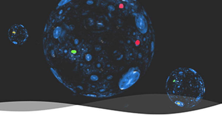
CUTTING EDGE TOOLSTO ACCELERATE MEDICAL RESEARCH

We are leaders in the development of RNA-focused research reagents.

About Lucerna

Founding Mission

The mission of Lucerna is to be the market leader in the development of RNA-based research reagents that enable scientific discovery.

Lucerna was launched in 2010 by scientists who developed the Spinach™ technology. Spinach, a RNA mimic of the green fluorescent protein (GFP), was developed in the laboratory of Dr. Samie Jaffrey of Weill Cornell University. Since then, over 50 articles have been published on the Spinach™ technology.

Lucerna licensed the Spinach™ technology from Cornell University with the goal of developing "scientists friendly" tools to help propel this burgeoning RNA era.

Product Launch

Lucerna's first RNA imaging product, DFHBI fluorophore, was launched in 2011.

DFHBI is a small molecule dye that fluoresces when bound to the Spinach aptamer. RNA that genetically encodes the Spinach tag can be expressed in cells and with the addition of DFHBI, living RNA localization and movements can now be visible under the microscope.

The development and testing of the Spinach aptamer and DFHBI fluorophore was first described in Paige, et al., Science, 2011.

New Vegetables

DFHBI-1T, a brighter and more photostable fluorophore, was launched in 2014. Soon after, new aptamer tags that have different properties or fluoresce in different colors (Broccoli, Corn, Red Broccoli, and Radish) were also introduced.

The Spinach aptamer was originally named as homage to the late Dr. Roger Tsien. Dr. Tsien named all of fluorescent proteins developed in his labs after fruits (e.g. mCherry, dTomato, mApple, mCitrine, etc.). Following this tradition, all aptamers derived from the Spinach™ technology are named after vegetables that best match their fluorescent colors.

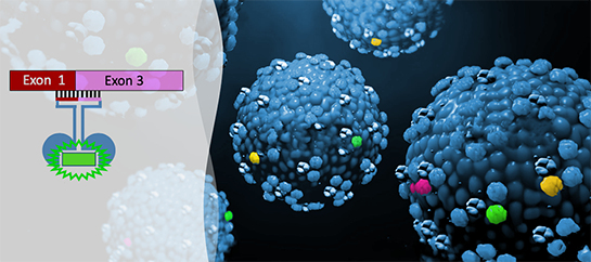
Splicing Drug Discovery Platform

Lucerna received a $1.5M phase II SBIR grant from the National Cancer Institute to develop a high-throughput drug screening platform to identify drugs that can correct a splicing event key to cancer's metabolic pathway. This platform uses the Spinach™ technology to generate fluorescent sensors that can detect endogenous splice isoforms in cells.

In 2017, Lucerna also successfully completed a R&D project for a top 5 pharma company to develop a custom splicing assay to identify novel splice modulating hits.

Our splicing drug discovery platform is high-throughput compatible, can target any endogenous spliced isoforms, and can detect hits that cause as small as 10% splicing changes.

In Development

Lucerna is always looking for ways to develop unique research and drug discovery tools to help solve urgent problems in the RNA world.

Current technologies in development include:
• Cell-based antiviral drug discovery platforms
• Enhanced fluorophores that allow single-molecule RNA imaging in live cells.
• Fluorescent sensors that report changes in RNA structures.
• Sensors that detect intracellular metabolic changes for drug discovery and for industrial biotechnology applications.

Products & Services

Fluorophores

Fluorophores

Proprietary dyes that turns on the fluorescence of the Spinach family of RNA aptamers.

MORE DETAILS
Assays

Assays

High-throughput compatible assays with homogenous workflows and robust fluorescence readouts.

MORE DETAILS
Sensors

Sensors

Custom fluorescent sensors that detect medically important RNAs with specificity and sensitivity.

MORE DETAILS

Featured Products

RNA Splice Detection Sensor Development

Service Quote Required

Custom fluorescent sensor platform for high-throughput drug discovery of RNA splice modulators.

Why RNA?

Published literature

    There are no papers matching your search terms.

    ALL PApers

    Team & Technology

    RNA plays a critical role in all aspect of life and errors in RNA cause diseases, many currently without cures. Therefore, it is the utmost important to have robust tools to see, study, and drug these RNAs.

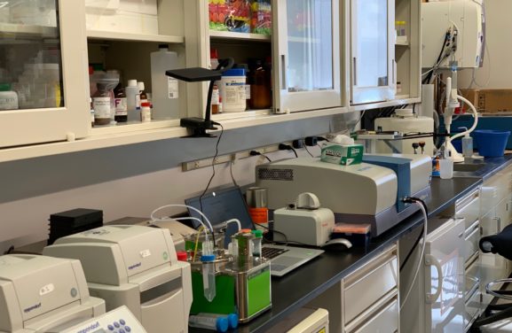
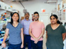
    Lucerna, Inc. is an early-stage biotech company comprised of high caliber scientists with extensive RNA biology and assay development background.  Located in the Downstate Biotechnology Center in Brooklyn, we are focused on developing and commercializing RNA-based research tools to help accelerate biological research and drug discovery.  At Lucerna, we strongly believe in combining the rigor of an academic research institution with the innovative spirit of a biotechnology company.  And also having fun while doing great science!

    Karen Wu

    Karen Wu President & Co-Founder

    Samie R. Jaffrey

    Samie R. Jaffrey Scientific Advisor, Co-Founder

    Jeremy S. Paige

    Jeremy S. Paige Co-Founder

    Carolyn E. Car

    Carolyn E. Car Cellular Assay Team Leader

    Ryan O'Hanlon

    Ryan O'Hanlon Splicing Assay Team Leader

    Aminoor Rashid

    Aminoor Rashid Machine Learning Team

    Shanai Brown

    Shanai Brown Splicing Assay Team

    Xinnan Guo

    Xinnan Guo Splicing Assay Team

    Arya Phatak

    Arya Phatak Cellular Assay Team

    Our Resources

    • News on the Spinach Technology

      The Scientist magazine compared and contrasted SpinachTM against the latest methods in tracking RNA in living cells.

      Media coverage on the SpinachTM technology by Science DailyPopular Science, and GEN.

      The Spinach aptamer was featured as the Molecule of the Month on the Protein Data Bank website.

      Spinach, Broccoli, and Corn aptamers were featured on the Addgene‘s blog.

      GEN reports the usage of the SpinachTM technology as metabolite sensors.

    • Publications about the Spinach technology

      Fluorophore-promoted RNA folding and photostability enable imaging of single Broccoli-tagged mRNAs in living mammalian cells.  Angewandte Chemie.  December 18, 2019.  LINK

      Highly efficient expression of circular RNA aptamers in cells using autocatalytic transcripts.  Nature Biotechnology.  June, 2019.  LINK

      Programmable RNA detection with a fluorescent RNA aptamer using optimized three-way junction formation.  RNA.  May 16, 2019.  LINK

      Spectral tuning by a single nucleotide controls the fluorescence properties of a fluorogenic aptamer.  ACS Biochemistry.  March 6, 2019.  LINK

      Detection of low-abundance metabolites in live cells using an RNA integrator.  Cell Chemical Biology.  February 14, 2019.  LINK

      RNA structures and cellular applications of fluorescent light-up aptamer.  Angewandte Chemie.  August 13, 2018.  LINK

      Genetically encoded catalytic hairpin assembly for sensitive RNA imaging in living cells.  Journal of the American Chemical Society.  June 26, 2018.  LINK

      Modular cell-internalizing aptamer nanostructure enables targeted delivery of large functional RNAs in cancer cell lines.  Nature Communications.  June 11, 2018.  LINK

      Development of genetically encodable FRET system using fluorescent RNA aptamers.  Nature Communications.  January 2, 2018.  LINK

      RNA-based fluorescent biosensors for detecting metabolites in vitro and in living cells.  Advances in Pharmacology.  October 25, 2017.  LINK

      Using a specific RNA-protein interaction to quench the fluorescent RNA Spinach.  ACS Chemical Biology.  October 23, 2017.  LINK

      Imaging RNA polymerase III transcription using a photostable RNA-fluorophore complex.  Nature Chemical Biology.  September 25, 2017.  LINK

      A homodimer interface without base pairs in an RNA mimic of red fluorescent protein.  Nature Chemical Biology.  September 25, 2017.  LINK

      A tale of two G-quadruplexes.  Nature Chemical Biology.  September 25, 2017.  LINK

      FASTmir: an RNA-based sensor for in vitro quantification and live-cell localization of small RNA.  Nucleic Acids Research.  June 6, 2017.  LINK

      A fluorescent split aptamer for visualizing RNA-RNA assembly in vivo.  ACS Synthetic Biology.  September 15, 2017.  LINK

      Sequence-specific biosensing of DNA target through relay PCR with small-molecule fluorophore.  ACS Chemical Biology.  May 9, 2016.  LINK

      Fluorescent RNA aptamers as a tool to study RNA-modifying enzymes.  Cell Chemical Biology.  March 17, 2016.  LINK

      A conformation-induced fluorescence method for microRNA detection.  Nucleic Acids Research.  March 6, 2016.  LINK

      Tandem Spinach array for mRNA imaging in living bacterial cells.  Scientific Reports.  November 27, 2015.  LINK

      Combining Spinach-tagged RNA and gene localization to image gene expression in living yeast.  Nature Communications.  November 19, 2015.  LINK

      In-gel imaging of RNA processing using Broccoli reveals optimal aptamer expression.  Chemistry & Biology.  May 21, 2015.  LINK

      Imaging metabolite dynamics in living cells using a Spinach-based riboswitch.  Proceedings of the National Academy of Sciences of the United States of America.  May 11, 2015.  LINK

      RNA signal amplifier circuit with integrated fluorescence output.  ACS Synthetic Biology  October 29, 2014.  LINK

      Broccoli: Rapid selection of an RNA mimic of green fluorescent protein by fluorescence-based selection and directed evolution.  Journal of the American Chemical Society.  October 22, 2014.  LINK

      Structural basis for activity of highly efficient RNA mimics of green fluorescent protein.  Nature Structural & Molecular Biology.  July 15, 2014.  LINK

      A G-quadruplex-containing RNA activates fluorescence in a GFP-like fluorophore.  Nature Chemical Biology.  June 22, 2014.  LINK

      Using Spinach-based sensors for fluorescence imaging of intracellular metabolites and proteins in living bacteria.  Nature Protocols.  January 9, 2014.  LINK

      Plug-and-play fluorophores extend the spectral properties of Spinach.  Journal of the American Chemical Society.  January 6, 2014.  LINK

      Understanding the Photophysics of the Spinach-DFHBI RNA Aptamer-Fluorogen Complex To Improve Live-Cell RNA Imaging.  Journal of the American Chemical Society.  December 18, 2013.  LINK

      E88, a new cyclic-di-GMP class I riboswitch aptamer from Clostridium tetani, has a similar fold to the prototypical class I riboswitch, Vc2, but differentially binds to c-di-GMP analogs.  Molecular BioSystems. December 18, 2013.  LINK

      Gene position more strongly influences cell-free protein expression from operons than T7 transcriptional promoter strength.  ACS Synthetic Biology.  November 27, 2013.  LINK

      A superfolding Spinach2 reveals the dynamic nature of trinucleotide repeat-containing RNA.  Nature Methods.  October 27, 2013.  LINK

      Unbiased Tracking of the Progression of mRNA and Protein Synthesis in Bulk and in Liposome-Confined Reactions.  ChemBioChem.  September 11, 2013.  LINK

      Universal aptamer-based real-time monitoring of enzymatic RNA synthesis.  Journal of the American Chemical Society.  September 18, 2013.  LINK

      Programmable folding of fusion RNA in vivo and in vitro driven by pRNA 3WJ motif of phi29 DNA packaging motor.  Nucleic Acids Research.  September 9, 2013.  LINK

      Universal aptamer-based real-time monitoring of enzymatic RNA synthesis.  Journal of the American Chemical Society.  August 30, 2013.  LINK

      The Spinach RNA Aptamer as a Characterization Tool for Synthetic Biology.  ACS Synthetic Biology.  August 30, 2013.  LINK

      New approaches for sensing metabolites and proteins in live cells using RNA.  Current Opinion in Chemical Biology.  August, 2013.  LINK

      Imaging bacterial protein expression using genetically encoded RNA sensors composed of RNA.  Nature Methods.  July 21, 2013.  LINK

      Designer nucleic acids to probe and program the cell.  Trends in Cell Biology.  December, 2012.  LINK

      Nanomolar fluorescent detection of c-di-GMP using a modular aptamer strategy.  Chemical Communications.  July 20, 2012.  LINK

      Fluorescence imaging of cellular metabolites with RNA.  Science.  March 9, 2012.  LINK

      RNA mimics of green fluorescent protein.  Science.  July 29, 2011.  LINK

    • Publications about the Diglycyl-Lysine antibodies

      (* Denotes that articled that used Lucerna’s diglycyl-lysine antibodies in the research.)


      * Profiling of ubiquitination modification sites in talin in colorectal carcinoma by mass spectrometry. Chemical Research in Chinese Universities. 2019. LINK

      Ubiquitin diGLY proteomics as an approach to identify and quantify the ubiquitin-modified proteome. The Ubiquitin Proteasome System. 2018. LINK

      * Examining ubiquitinated peptide enrichment efficiency through an epitope labeled protein. Analytical Biochemistry. 2016. LINK

      * System-wide analysis of BCR signalosomes and downstream phosphorylation and ubiquitylation. Molecular Systems Biology. 2015. LINK

      * Profiling lysine ubiquitination by selective enrichment of ubiquitin remnant-containing peptides. Exocytosis and Endocytosis. 2014. LINK

      Global ubiquitination analysis by SILAC in mammalian cells. Stable Isotope Labeling by Amino Acids in Cell Culture (SILAC). 2014. LINK

      Comprehensive profiling of protein ubiquitination for drug discovery.  Current Pharmaceutical Design.  2013.  LINK

      * A mental retardation-linked nonsense mutation in cereblon is rescued by proteasome inhibition.  Journal of Biology Chemistry.  2013.   LINK

      * Systems-wide analysis of ubiquitylation dynamics reveals a key role for PAF15 ubiquitylation in DNA-damage bypass.  Nature Cell Biology.  2012.  LINK

      * Proteomic analyses reveal divergent ubiquitylation site patterns in murine tissues.  Molecular & Cellular Proteomics.  2012.  LINK

      * Synaptic protein ubiquitination in rat brain revealed by antibody-based ubiquitome analysis.  Journal of Proteome Research.  2012. LINK

      The new landscape of protein ubiquitination.  Nature  Biotechnology.  2011.  LINK

      Global identification of modular Cullin-RING ligase substrates.  Cell.  2011.  LINK

      * A proteome-wide, quantitative survey of in vivo ubiquitylation sites reveals widespread regulatory roles. Molecular & Cellular Proteomics.  2011.  LINK

      * Global analysis of lysine ubiquitination by ubiquitin remnant immunoaffinity profiling.  Nature  Biotechnology.  2010.  LINK

    • Posters & Presentations

      Overall view of Lucerna technology platform. Download

      Tools for splice modulation drug discovery: SpinachTM splice sensor platform. Download

      New cell-based high-throughput screening platform for RNA-mediated neurodegenerative diseases. Download

      Cyclic-di-GMP assay kit: A first-in-class HTS assay for bacterial tolerance drug discovery. Download

      HTS platforms for diverse RNA-targeted drug discovery applications. Download


    • RNA imaging in bacterial cells

      E. coli cell imaging protocol

      1. Transform BL21 Star (DE3) or similar E. coli competent cells with 40 ng of plasmid DNA expressing the tRNA-Spinach chimeras in a bacterial expression vector under the control of a T7 promoter (e.g. pET28c).

      2. After overnight growth, pick single colonies for inoculation in Luria Broth containing kanamycin (LB-Kan).

      3. At OD600 = 0.4, induce culture with 1 mM of IPTG and continue shaking at 37 ºC for more 2 hours.

      4. During this time, coat glass-bottom dishes with 0.1 mg/mL poly-L-lysine (PLL) for at least 2 hours in 37 ºC. Wash the dishes at least twice with ddH2O to remove free PLL. (Dishes can also be coated overnight at 4 ºC)

      5. Remove and spin down 100 µL of culture, then resuspend in 2 mL of pH 6.0 M9 minimal media.

      6. Plate 200 µl aliquot of resuspended culture on poly-L-lysine (PLL)-coated glass-bottom dishes and incubate for 45 minutes at 37o C.

      7. Wash adherent cells twice with M9 media and then incubate with 200 µM DFHBI in pH 6.0 M9 minimal media for 45 min at 37o C.

      8. Take live fluorescence images with a 60X oil objective using a FITC filter.


      M9 minimal media:

      1X M9 salts

      2 mM MgSO4

      0.1 mM CaCl2

      0.4% glucose


      5X M9 salts:

      64 g NaHPO4 • 7H20

      15 g KH2PO4

      2.5 g NaCl

      5 g NH4Cl

      Add water to 1 L and autoclave

    • RNA imaging in mammalian cells

      HEK293T cell imaging protocol

      (This protocol can be used to image RNAs tagged with any of Lucerna’s fluorogenic RNA in common mammalian cell lines)

      1. Coat glass-bottom dishes (Mattek #P24G-1.5-13-F) with 100 µg/mL of poly-L-lysine (PLL) overnight at 4°C.

      2. Wash the dishes twice with ddH2O and UV sterilize for 5 min. Coat the sterilized dishes with 100 µg/mL rat collagen-I and 50 µg/mL laminin for 2 hours at 37°C.

      3. After removing the coating solution and allowing the dishes to air-dry, plate HEK293T cells at 80,000 cells/ml.

      4. After 24 hours, transfect HEK293T cells with 0.4 μg of Spinach2-tagged RNA construct with FuGene HD [0.4 µg DNA, 1.8 µL FuGene HD (Promega #E2311), in Opti-MEM => 20 µL/well]. Imaging experiments are typically performed 48 hours after transfection.

      5. Thirty minutes prior to experiment, replace HEK293T cell media with imaging media (DMEM with no phenol red or vitamins and supplemented with 25 mM HEPES, 5 mM MgSO4, and 5 μg/ml Hoechst 33342) and 20 μM DFHBI-1T or vehicle are added to the cells to promote RNA-fluorophore complex formation.

      6. Live fluorescence images of HEK293T cells are taken with the DIC (Normal) microscope using the 63X oil objective.

      7. DFHBI-1T fluorescence are detected using the FITC filter (~100 ms exposure), Hoescht 33342 fluorescence with the DAPI filter (~100 ms exposure,) and phase contrast images are taken at ~500 ms.

      8. To remove any green autofluorescence or residual fluorescence from DFHBI-1T, take images of untransfected control cells with DFHBI-1T and subtract this background fluorescence from Spinach2-containing cells.

      Note: Unless specifically stated, all reagents were purchased from ThermoFisher.

    • Selective enrichment of ubiquitin remnant peptides

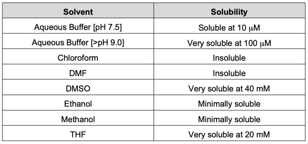
    Questions on DFHBI and DFHBI-1T fluorophores:

    • What is the solubility of DFHBI and DFHBI-1T fluorophore in commonly used solvents?

    • What is the recommended protocol to test DFHBI and DFHBI-1T fluorescence?

      For all our DFHBI and DFHBI-1T batches, we can verify fluorescence with Spinach2™ or Broccoli™ aptamer alone in vitro. The protocol is as follows:

      1a. PCR Spinach2™ sequence with T7 binding site. Gel-purify the 114 bp product.

      Spinach2™ sequence:
      GATGTAACTGAATGAAATGGTGAAGGACGGGTCCAGTAGGCTGCTTCGGCAGCCTACTTGTTGAGTAGAGTGTGAGCTCCGTAACTAGTTACATC

      T7-Spinach2™ 5′: taatacgactcactatagg GATGTAACTGAATGAAATGGTGAAGGACG
      Spinach2™ 3: GATGTAACTAGTTACGGAGCTCACACTC
      (Anneals at 55 °C)

      1b. PCR Broccoli™ sequence with T7 binding site. Gel-purify the 49 bp product.

      Broccoli™ sequence:
      GAGACGGTCGGGTCCAGATATTCGTATCTGTCGAGTAGAGTGTGGGCTC

      T7-Broccoli™ 5′: taatacgactcactatagg GAGCCCACACTCTACTCG
      Broccoli™ 3: GAGCCCACACTCTACTCG
      (Anneals at 50 °C)

      2. Assemble in vitro transcription reaction using any commercial T7 in vitro transcription kit per manufacturer protocol with 100 ng PCR template. Incubate in 42 °C water bath overnight.

      3. Treat with 1 µL DNase for 15 min at 37 °C. Purify by phenol:chloroform extraction and isopropanolol precipitation (as described in the T7 in vitro transcription kit manual). Resuspend RNA in water.

      Alternately, Bio-Rad Micro Bio-spin P30 gel columns can be used to purify and concentrate RNA.

      4. Make 5X aptamer buffer: 0.75 M KCl, 200 mM HEPES, and 0.5 mM MgCl2

      5. Dilute 1  µM RNA in 1X aptamer buffer with or without 20 µM DFHBI. In a spectrofluorimeter, excite the solution at 447 nm (for DFHBI) and 482 nm (for DFHBI-1T) and measure fluorescence emission from 495-600 nm.

    Questions on Diglycyl-Lysine Antibody:

    • How much diglycyl-lysine antibodies and lysates should be used in an ubiquitin remnant pull-down experiment?

      In our hands, we typically use 100 micrograms antibodies coupled to 20 microlitter Affigel 10 beads and 10 – 100 micrograms of purified ubiquitinated proteins from transfected cells for a pull-down experiment. Similarly, the recently Weinert et al. paper, which cited the use of our antibodies, used ~20 milligram proteins from non-transfected cells and ~100 microgram antibody coupled to protein G beads for immunoprecipitation. So 10-100 micrograms of proteins would be the suitable range.

    • What is the recommended dilution for performing western blot analysis using the diglycyl-lysine antibody (GX41)?

      For the western blot showed in the product data sheet, we used the GX41 antibody at 1:500 dilution. However, in these experiments we validate the specificity of the GX41 antibody against chemically Gly-Gly modified brain lysates and large peptides (b-lactoglobulin and lysozyme).

      We don’t recommend doing western blot for ubiquitinated cellular proteins with the GX41 antibody because the transfer of trypsinized peptides on to membrane are incredibly inefficient due to the small size of the peptides. This problem can be some what mitigated by either putting a
      second membrane while transferring to catch any peptides that get through the first membrane or use a membrane with size cut-off that is smaller than your peptides of interest. Additionally, you can also increase the loading protein concentration to ensure more peptides are transferred onto the blot.

      The most sensitive method to assay for ubiquitinated proteins are to IP with GX41-conjugated resins and mass spec the captured peptides. A complete protocol on how to perform pull-down experiment with GX41 antibody can be found in the Protocols section.

    • What is the explanation for the occasional presence of precipitates in the antibody solution after resuspension?

      Due to the process of lyophilization, there can be some undissolved salts that appear as precipitates. We always pack each tube with extra antibodies to compensate for possible antibody loss during resuspension. If you need to determine the actual concentration, we recommend that you spin down the precipitates and determine the final concentrations of the resuspended antibodies by performing the Bradford assay with IgG or BSA standard curve.

    • Which solid support is recommended for diglycyl-lysine antibody conjugation?

      The efficient conjugation of diglycyl-lysine antibodies to resins is crucial to the success of the pull-down experiments. Therefore, we recommend that you use Affi-gel resins, as stated in the Xu et al Nature Biotechnology paper. In our hands, we found that Affi-gel resins gave the best coupling while protein A/G beads often showed at least 50% reduction in coupling efficiency. Also, the antibody concentration must be above 1.0 mg/ml. If you are using small amounts of antibodies then adjust the volume appropriately to get the desired concentration. A complete protocol on how to conjugate GX41 antibody to affi-gel resins can be found in the Protocols section.

    • Databases

      Circular RNA (circRNA):

      • circBase – Public circular RNA datasets
      • circ2Traits – A comprehensive database for circular RNA potentially associated with disease and traits


      Exosome:

      • ExoCarta – An exosome database that provides with the contents that were identified in exosomes in multiple organisms
      • exoRBase – A repository of circular RNA, long non-coding RNA and messenger RNA  derived from RNA-seq data analyses of human blood exosomes


      Extracellular RNA (exRNA):

      • exRNA – A research portal for all publications, resources, standards generated from NIH Common Fund-supported Extracellular RNA Communication program
      • exRNA atlas – The data repository of the Extracellular RNA Communication Consortium (ERCC)


      MicroRNA (miRNA):

      • Cupid – A method for simultaneous prediction of miRNA-target interactions and their mediated competitive endogenous RNA interactions
      • miRBase – A searchable database of published miRNA sequences and annotation
      • TargetScan – Predicts biological targets of miRNAs by searching for the presence of conversed sites that match the seed region of each miRNA


      Non-Coding RNA (ncRNA):

      • LNCipedia – A comprehensive compendium of human long non-coding RNAs
      • NONCODE – An integrated knowledge database dedicated to non-coding RNAs (excluding tRNAs and rRNAs)
      • RNAcentral – The non-coding RNA sequence database


      Proteins:

      • FPbase – Fluorescent protein database
      • PDB – A repository of information about the 3D structures of proteins, nucleic acids, and complex assembles


      RNA Modification:


      Other:

      • Apta-Index™ – A catalog of published aptamers 
      • LncRRIsearch – A web server for lncRNA-RNA interaction prediction integrated with tissue-specific expression and subcellular localization data
      • PRD – A protein-RNA interaction database
      • Rfam – A collection of RNA families, each represented by multiple sequence alignments, consensus secondary structures and covariance models
      • RPISeq – A family of machine learning classifiers for predicting RNA-protein interactions using only sequence information.


      Small nucleolar RNA (snoRNA):

      • Plant snoRNA database – A database of Arabidopsis snoRNA
      • snoRNABase – A comprehensive database of human H/ACA and C/D box snoRNAs
      • snoRNAdb – An algorithm for searching genomic sequence for 2′-O-ribose methylation guide snoRNA genes


      Transfer RNA (tRNA):

      • mitotRNAdb – A database of mitochondrial tRNA genes
      • tRNAdb – Compilation of tRNA sequences and tRNA genes


      (Please reference the appropriate papers when using these databases)

    • Games with purpose

      EteRNA – An online game that engage players to solve puzzles related to the folding of real-life RNA molecules whose structures may or may not been known

      FoldIt – An online game that allow players to design new or improved protein structures for the purpose of advancing our understanding of protein folding and creating new therapeutics

      Phylo -An online game that let players optimize multiple sequence alignments of different phylogentic taxa in order to identify functional sites, uncover mutation events, and trace the source of genetic diseases

    • RNA structure prediction softwares

      KineFold – RNA/DNA folding predictions including pseudoknots and entangled helices

      UNAFold – Web-based services for nucleic acid fold and hybridization predictions

      NUPACK – Software suite for the analysis and design of nucleic acid structures, devices, and systems developed

      OxDNA –  Coarse-grained models that simulate the thermodynamic and mechanical properties of single- and double-stranded DNA and RNA

      RNAstructure – Programs and web services developed in Dr. David Matthew’s lab at the University of Rochester.  Algorithms include:

      • biFold – Predict the lowest free energy structure for two interacting sequences, allowing intramolecular base pairs
      • DuplexFold – Predict the lowest free energy structure for two interacting sequences, not allowing intramolecular base pairs
      • EnsembleEnergy – Calculate the ensemble folding free energy change for a sequence
      • PARTS – Predict the common secondary structure, including base pair probabilities, for two unaligned sequences
      • MaxExpect – Generate a structure or structures composed of highly probable base pairs
      • RNAbows – Visualize partition function calculations of base pairing probabilities
      • Stochastic – Generate a representative sample of structures using stochastic sampling


      ViennaRNA – Programs and web services developed by the University of Vienna. Algorithms include:

      • RNAalifold – Predicts consensus secondary structures from an alignment of several related RNA or DNA sequences
      • RNAfold – Predict secondary structures of single stranded RNA or DNA sequences
      • RNAinverse – Allows you to design RNA sequences for any desired target secondary structure
      • RNAprobing – Predicts minimum free energy structures and base pair probabilities from single RNAs using a guiding potential based on SHAPE reactivity probing data


      (Please reference the appropriate papers when using these programs)

    • Useful sites

      BioMath Calculator – DNA, RNA, and protein conversions; dilution and molarity calculators provided by Promega

      Journal of Visualized Experiments (JOVE) – Videos for many detailed protocols for advanced techniques

      NEB tools – Useful tools and resources provided by NEB

      OligoCalc – Oligonucleotide property calculator

      Useful numbers for biologists – The database for useful biological numbers

    Get In Touch

    Drop Us A Line