Products
BI
Information

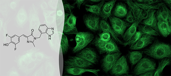
(Z)-3-((1H-benzo[d]imadazol-4-yl)methyl)-5-(3,5-difluoro-4-hydroxybenzylidene)-2-methyl-3,5-dihydro-4H-imidazol-4-one (BI)
MOLECULAR FORMULA:
C19H14F2N4O2
MOLECULAR WEIGHT:
368.3
CAS NAME/NUMBER:
Fluorescence Spectra
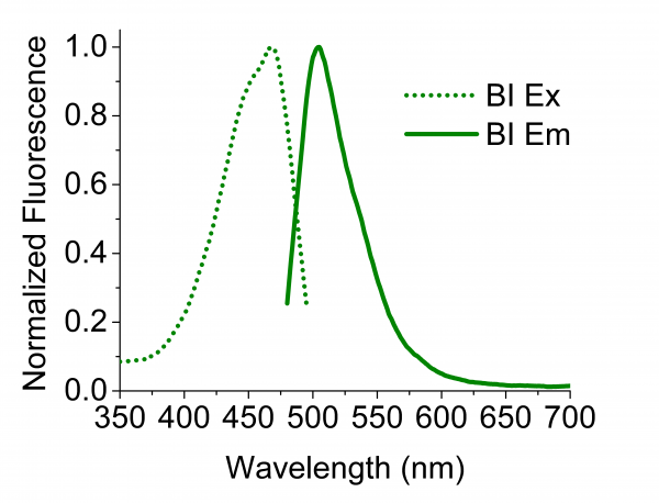
Absorption and fluorescence emission spectra of BroccoliTM/BI in pH 7.4 buffer.
PRESENTATION: Lyophilized dye
QUALITY ASSURANCE: Products are analyzed by 1H NMR and LC-MS and provided at purity of >98% by HPLC.
USAGE STATEMENT: This product is intended for research use only and are not to be used for any other purpose, which includes but is not limited to, unauthorized commercial uses, in vitro diagnostic uses, ex vivo or in vivo therapeutic uses or any type of consumption or application to humans or animals. Due to the highly specific nature of fluorophores and aptamers, we cannot predict or be held responsible with respect to how LucernaTM products will behave in its customers’ systems. Researchers using LucernaTM products should conduct optimization studies to achieve the optimal result possible for their intended application.
Data
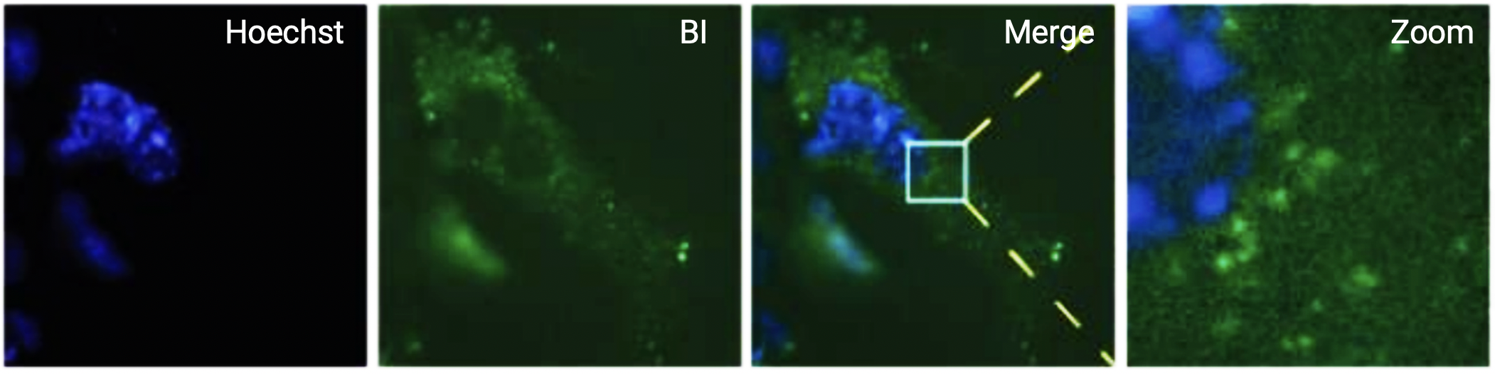
Figure 1. Live-cell imaging of COS7 cells expressing β-actin mRNA transcripts tagged with 24 copies of BroccoliTM in the 3’UTR. Cells were incubated with 0.5 µg/ml Hoechst 33342 stain (blue) and 10 µM BI (green) and images were acquired using a 500 msec exposure with a GFP filter set. Mobile fluorescence puncta were observed only when Broccoli-expressing cells were treated with BI.
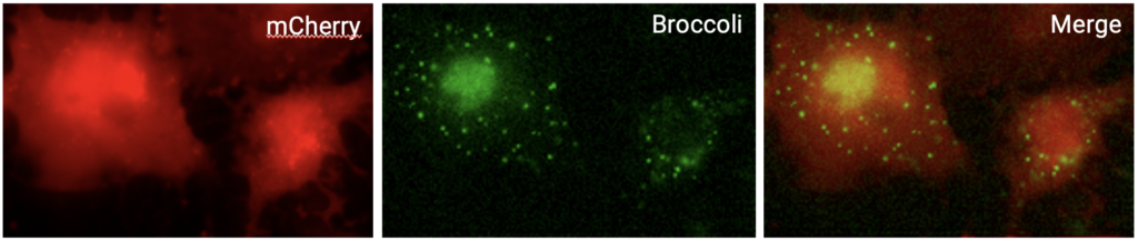
Figure 2. For single-molecule imaging, COS7 cells were transfected with a construct expressing mCherry-24xBroccoliTM transcripts and imaged in the presence 10 µM BI. Images of the same cells were acquired using TRITC and GFP filter sets to capture mCherry fluorescence (red) and BroccoliTM (green) signals. Mobile fluorescent puncta were detected in both the nuclei and cytosol of mCherry-expressing cells.

Figure 3. Live fluorescent mRNA puncta were imaged using COS7 cells expressing either A. the MS2-GFP system and B. the BroccoliTM-BI system. The puncta size of Broccoil-tagged mRNA is similar to the puncta size of MS-tagged mRNA. Further, the puncta intensities of mRNA labeled from both systems exhibit a single Guassian distribution, consistent with the majority of these puncta reflecting single mRNA transcripts. The scale bars, are both 5 µm.
Reference
Fluorophore-promoted RNA folding and photostability enable imaging of single Broccoli-tagged mRNAs in living mammalian cells. Li X, Kim HY, Litke JL, Wu JH, Jaffrey SR. Angew Chem Int Ed. 2020 Mar 9;59(11): 4511-4518.